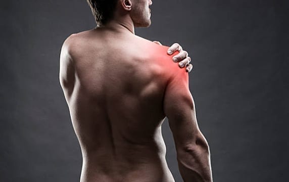
Shoulder Injury Understanding Shoulder Injuries
Shoulder injuries are typically caused by athletic activities that involve excessive, repetitive, overhead motion, such as swimming, tennis, pitching, and weightlifting. Injuries can also occur during day-to-day activities such as: washing the car, hanging curtains, and gardening.
These are three common shoulder injuries caused by sports participation:
Superior Labral Tear - aka SLAP Tear
A common diagnosis for shoulder injuries is a superior labral tear, aka SLAP tear. SLAP stands for Superior Labral tear Anterior to Posterior. There many different types of SLAP tears, with different variations of severity and treatments.
Q: What is a SLAP Tear?
A: A tear to the ring of cartilage around the shoulder socket. A SLAP tear occurs over time from repetitive, overhead motions, such as throwing a baseball, playing tennis or volleyball, or swimming.
Common symptoms:
Q: Sounds you may notice if you have a SLAP tear?
A: Clicking, grinding, locking, and/or popping.
Labral tears are basically cartilage tears from the glenoid soffit of the glenohumeral joint and are involved in shoulder instability. Classically this is seen with the anterior labrum with a shoulder dislocation. These typically present with a first time dislocator, although 2% of the time the labrum is not involved and either can be a capsular stretch or a rupture of the capsule as well. But in most instances it involved the labrum. Once the shoulder is located we evaluate the patient, their demand level needs and their age, and discuss treatment options. The risks of recurrence of labral tears is higher amongst younger patients if they are conservatively treated, and most data suggests that operative intervention is the best way to treat initial dislocators, and definitely recurrent dislocators. When we look at labral tears we want to make sure that the patient does not have any other associated injuries because these are usually high level injuries, such as a nerve injury.

So a careful evaluation involving the neurovascular structures is performed. The patient will be checked for their range of motion. They will also be checked to see if they have an apprehension sign, which means at the 90/90 position the patient will show apprehension because the labrum is lost as a passive checkrein mechanism to locate the shoulder in that position. So that can be relieved with a relocation sign with the patient lying down, and the humeral head is pushed down to be relocated. With respect to shoulder instability, it can be graded. Usually labral tears in the anterior quadrant are unidirectional, but there can be a component of multidirectional instability. . So we look for other signs like a Sulcus sign, hypermobility, but ultimately when the decision has been made for surgery and examination under anesthesia is performed on that shoulder to see if there are other signs of instability besides unidirectional instability in the classic anterior labral tear. There can be SLAP tears, which is a superior labrum anterior posterior, which is not as dramatic as an anterior inferior labral tear.
They do not demonstrate the same instability issues because the anterior inferior glenohumeral ligament is attached to the anterior inferior labrum, and that is a key structure that is a checkrein against anterior translation of the humeral head in the 90/90 position. However, SLAP tears can be symptomatic. On physical exam they can be positive with an O'Brien's test. They can also have other physical findings such as a relocation and an apprehension sign as well if there is a significant anterior superior component. Posterior labral tears also exist and can be picked up on physical exam as well with a load and shift posteriorly. Labrums that are symptomatic are typically fixed arthroscopically with arthroscopic techniques. That procedure could take 15 to 20 minutes arthroscopically. Recovery although is longer. The patients are in a sling for six weeks, then they start physical therapy. They are usually in therapy for three to six months before full recovery. Also if there is associated stretch with the capsule, a capsulorrhaphy can be performed at the same time. That is basically sewing the capsule and tightening the capsule to provide more stability to the shoulder.

Shoulder Instability
Athletes commonly experience Shoulder Instability. This injury often occurs when you’re partaking in contact sports (football, hockey, or ones that require repetitive movements, like baseball).
Q: How does Shoulder Instability happen?
A: Shoulder instability happens when your ligaments, muscles, and tendons no longer secure your shoulder joint. Shoulder instability results in, the humeral head (circular, top part of your upper arm bone) to be dislocated.
Q: What is a Dislocation?
A: A dislocation is characterized by severe, abrupt onset of pain; subluxation (partial dislocation) may be accompanied by short spurts of pain. Other symptoms may include: weakness in the arm, lack of motion. Swelling and bruising on your arm are visible changes you may also notice.
Rotator Cuff Injury
Rotator cuff injuries are very common. Each year, approximately 200,000 Americans require shoulder surgery to repair a torn rotator cuff.
Q: What is a Rotator Cuff Injury?
A: This is another injury commonly seen in players participating in repetitive, overhead sports. Some of these sports include: swimming, tennis, and baseball. Rotator cuff injuries are typically categorized by weakness in the shoulder, reduced range of motion, and stiffness.
Rotator cuff injuries are also painful. Here’s what you need to know:
What are some common symptoms related to Rotator Cuff Tears injuries:
Like to a SLAP tear, people with rotator cuff injuries often experience achy shoulder pain.
Early treatment is the key to the best possible outcome regarding full recovery!
Rotator cuff tears are the most common entity that we treat in this office. We treat a lot of patients with work related injuries or accidents or athletes both amateur and professional with this problem. Rotator cuff tears are important because they basically effect the way the shoulder functions, and it is a very important structure because it smooths out the movement of the very large deltoid muscle, and it depresses the humeral head, and it dynamically stabilizes the should joint. So when it is injured patients really have a decrease in shoulder function and oftentimes complain of constant pain with and without movement. A lot of times patients with full thickness rotator cuff tears will not be able to lift their arm up against gravity or have profound weakness again gravity. Patients are typically in their 5th, 6th or 7th decade, but can be younger depending on the mechanism of injury.

Typically it is an eccentric load to the shoulder where a force overwhelms the shoulder joint and as a result the tendon gets overloaded and tears. Tears can either be partial or full thickness. Partial tears also present the same way with pain with pain and the inability to lift the arm. The physical examination with patients that have rotator cuff tears, one of the hallmarks is as, meaning the scapula kicks out. They get some winging and the smoothness of movement is lost in comparison to the non injured shoulder, and both shoulders should be examined. Oftentimes they have pain as the arm gets higher in the air. Sometimes they cannot get past a certain amount of what we call forward elevation. Another hallmark of physical exam is muscle testing, and we are able on physical exam to isolate the shoulder rotator cuff tendon and see if it can sustain itself against gravity or against external rotation. When we note weakness then we are suspicious of a rotator cuff tear. The next evaluation in terms of the rotator cuff tendon would be palpation of the structure, and this is done with patient seated. The arm is placed in extension and external rotation and the rotator cuff is palpated, and if tenderness is elicited by bringing it into extension and then abduction and external rotation, then that could be positive also in terms of physical finding for a rotator cuff tear.
We also test the Napoleon sign, or belly press sign, for the subscapularis tendon or the lift-off sign as well. We look for other pain generators in addition to the rotator cuff tendon, but that will be discussed in a later section. We obtain appropriate x-rays to make sure there is no other disease entities and to make sure that the humeral head is depressed on the Grashey view and there is no humeral head elevation. We also look for acromial morphology and glenohumeral arthritis as well. The next issue in terms of the workup would be the MRI. We usually get an MRI without contrast. The MRI is valuable because it can give us the information as to whether it is a full thickness rotator cuff tear or a partial thickness rotator cuff tear. It can tell us how far the tendon is retracted. It can also tell us whether there is fatty substitution in the muscle, an indication of a chronic tear. It can give us some prognostic indicators preoperatively as well depending on the quality of the MRI. It will also qualify the extent of the tear as well and other associated pathologies within the shoulder. So it is an important part of the workup. If there is a partial thickness tear, and the patient has good rotator cuff strength, then a physical therapy program would be the first line of treatment.

And if physical therapy failed, then injections would be performed. If the injections failed and a total of three months of therapy minimally has been done and the patient has not been improved then at that point arthroscopic surgery can be considered to go in and perhaps do an arthroscopic repair of that partial tear and a subacromial decompression and address other associated pain generators. With respect to full thickness tears, that would be addressed with an arthroscopic repair as well. Typically that is done as an outpatient procedure. The technique that we use, using the SpeedScrew, allows us to intraoperatively individually tension each anchor point so as to maximally load the tendon and compress it within the footprint, yet at the same time allow for medial movement and sliding of the tendons interdigitations which is a huge advantage over other current treatment methods that employ a medial row of an anchor that captures the tendon and prevents functioning of the rotator cuff. It is important to note that medial row repairs have a high rate of retear, 20% to 30%, and more than half of those are medial ruptures at the muscle tendon junction for which there is no treatment.
So our technique, in my opinion, is a huge advantage over that, and yet at the same time our technique allows us to restore the footprint and maintain footprint mechanics anatomically. This allows the ultrastructural sliding of the supraspinatus and infraspinatus interdigitation and does not compromise muscle tendon junction at all. We do our surgeries as an outpatient because it is arthroscopic. The portals are small. Procedure time is usually about 30 to 40
minutes, and the patients go home the same day. We start passive range of motion in physical therapy right away, and they get to do passive range of motion for six weeks. Then after six weeks they are doing active range of motion, which is strengthening. By about three to four months patients are done with their therapy program and are allowed to basically return to activities of daily living.
Biceps Problems
The next problem is biceps problems. The long head of the biceps has been implicated in a lot of pain generation. Typically it is found in conjunction with other pathologies. The best way to determine on physical exam is basically palpating the structure, the long head of the biceps, and if it produces pain then there is bicipital tendinitis.
You may or may not see it on an MRI. Injections can help in terms of making the diagnosis. With respect to treatment, physical therapy rarely is helpful in the treatment of biceps problems. In the event that conservative management has failed, arthroscopy, a biceps tenotomy or cutting of the biceps tendon would be performed.
Then a small open incision where a tenodesis of the biceps would be done where the new length tension relationship of the biceps would be established with coning out a small hole in the humerus and using a BioScrew to fix the biceps into the humeral head. Biceps tenotomies are also acceptable in terms of treatment of biceps problems, not fixing the biceps, just cutting the long head of the tendon. The problem is there can be spasm of the muscle, and that can occur in about 20% of the patients. So, I prefer to do an open subpectoral tenodesis in these issues.
AC Joint Arthritis
Arthritis of the AC joint is also a problem. Typically patients have pain over the AC joint based on palpation over that structure. The AC joint can be injected oftentimes, and once injections fail then the treatment would then be arthroscopic.
We do an arthroscopic resection of the clavicle, anywhere from 5 mm to 10 mm, but we try to err toward 5-6 mm. Less is more. Once you get more than 10 mm you risk the problem of instability of the AC joint ligament by damaging them, and that can be worse than the original pain. So, we take great care to not take more than 6 mm of distal clavicle. We focus on the posterior inferior spur because that is the one that is the most symptomatic.

Arthritis Of The Shoulder
The shoulder joint is a nonweightbearing joint. It basically can have arthritis, but arthritis is better tolerated in the shoulder than it is in the hip and the knee. So conservative management is paramount. You can do injections.
There are several off-label injections that we do in this practice including the Regenokine Program. However, if operative treatment is elected I like to use a stemless total shoulder, or a hemiarthroplasty where we only address the defect and the glenohumeral component. Rarely do we do an inlay glenoid. Compared to an anatomic total shoulder, the inlay hemiarthroplasty, the stemless procedure, has less blood loss. It takes about 40 minutes to do and is superior in range of motion and return to function.
Shoulder Impingement
Shoulder impingement, which is basically inflammation of the rotator cuff, is also treated. Typically those patients may have overused the shoulder. They present with rotator cuff symptoms because it is inflammation of the rotator cuff, but at an earlier stage.This is most often treated with physical therapy and conservative management and injections. Then if conservative management does not work, then we can go ahead and do an arthroscopic subacromial decompression.
